Madhubala, one of Bollywood’s most enigmatic actors, died at a rather young age due to a congenital heart defect known as ventricular septal defect (VSD). Born as a ‘blue baby’, she had a hole in her heart wall that separates the right and the left ventricles. This causes the blood to ‘leak’ from one ventricle to another and the complications that result from this can be life-threatening, if it is not treated in time.
While the field of medicine was not advanced enough to save Madhubala’s life, the condition can be treated successfully today in most children. Commonly known as ‘hole in the heart’, VSD occurs in 0.1 to 0.4 per cent of all live births and makes up about 20 to 30 per cent of congenital heart lesions and is one of the most common congenital heart diseases.
The heart’s structure
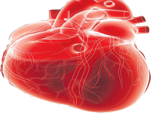
To understand the defect and its causes, symptoms and treatment, it is important to understand the heart’s structure. The human heart has four chambers: the right and left upper chambers are the atrium, while the right and left lower chambers are the ventricles. The presence of septum separates the heart into two halves, the right and left chambers. The septum dividing the upper chamber, that is the right atrium from the left
atrium, it is known as atrial septum, while the septum dividing the lower chamber, that is, right ventricle from the left ventricle, is known as ventricular septum.
The right and left ventricles of the heart are not separate before the baby is born. As the foetus grows inside the womb, a wall forms to separate these two ventricles. In cases where the wall does not completely form due to a defect, a hole remains. This hole is known as a ventricular septal defect. It is mostly a birth defect (congenital).
As with many other congenital diseases, the exact cause of the defect is not yet known. However, it is believed that the congenital heart defect results from problems early in the heart’s development. Environmental factors as well as genetics are considered to play a role in the occurrence of VSD. In some cases, VSD occurs by itself, while in others, it occurs in conjunction with other congenital diseases.
Size and location
The location and size of the VSD in the septum determines, in part, the consequences. If it is small in size, it rarely causes any problem. Small VSDs are recognised by a murmur, an extra heart sound heard via stethoscope during a regular physical checkup. As it does not put any pressure on the heart or lungs, it does not show any noticeable symptoms.
However, if the VSD is larger, the scenario is very different. During ventricular contraction, some of the blood from the left ventricle leaks into the right ventricle. This blood in turn goes to the lungs and re-enters the left ventricle via the pulmonary veins and left atrium. This complex flow of blood to and fro from the ‘hole’ causes a volume overload in the left ventricle and leads to pulmonary hypertension as well.
Therefore, if the hole is large, too much blood will be pumped to the lungs, and may cause heart failure. In such cases, blood leaks from one ventricle to another and eventually to the lungs; extra blood is pumped into the lung arteries making the heart and lungs work harder. The consequent congestion in lungs may also cause permanent
damage of the organs. Due to this, the child tends to breathe faster and harder than normal, while infants may face problem while feeding and growing at a normal rate.
Treatment options
While a small-sized VSD can be treated with minor surgeries and treatment, a large-sized VSD often needs open-heart surgery to close the hole. Closing a large VSD by open-heart surgery usually is done in infancy or childhood. It is also done in patients with few symptoms so that future complications can be prevented. Over the decades, diagnosis and treatment for VSD has improved.
Today, medical developments have enabled the successful closure of the hole, helping the child grow up without any complications and become a productive adult.
(The author is head and director, cardiology, Paras Hospitals, Gurgaon)
